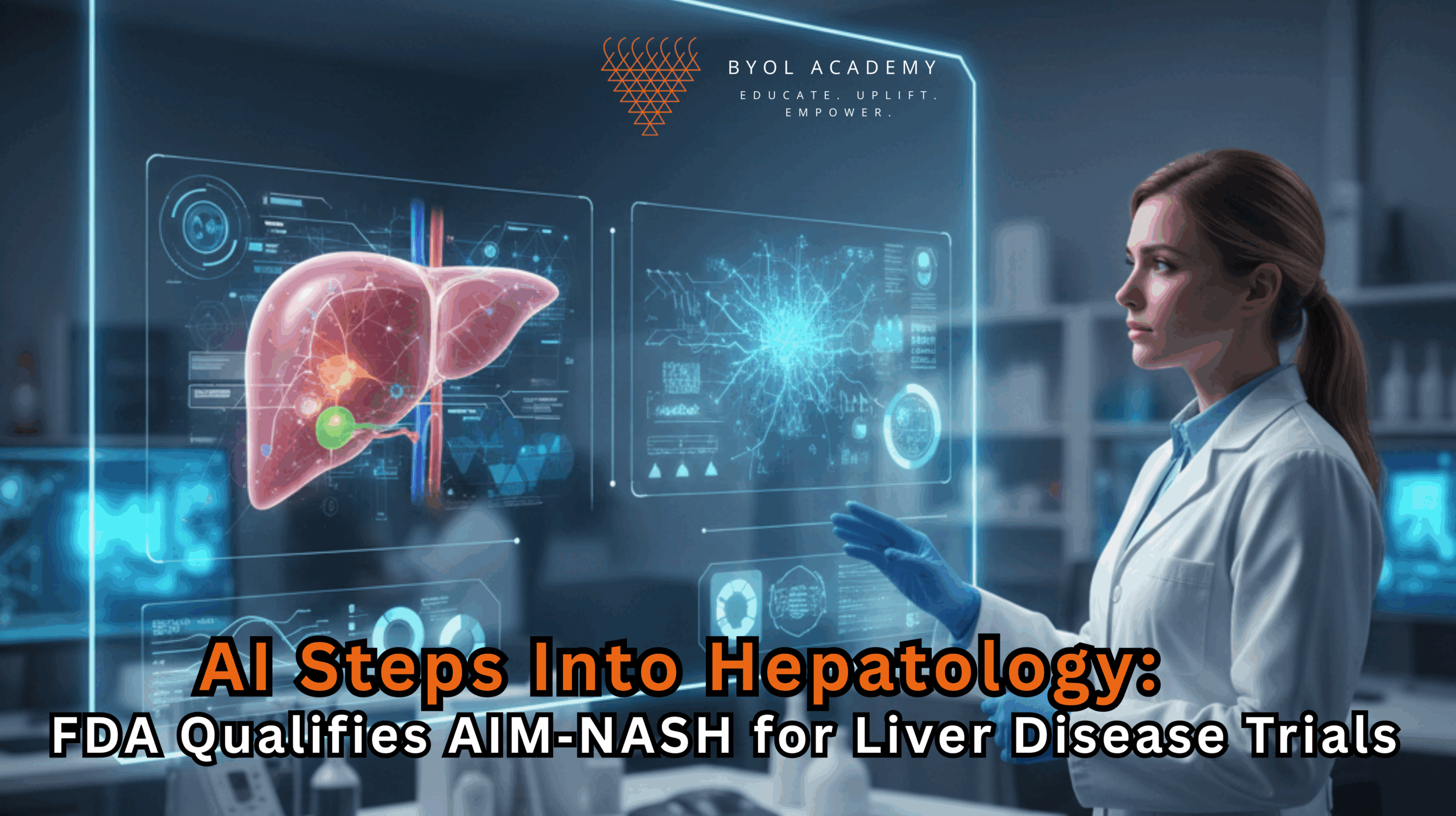
Recent developments in regulatory science have marked a pivotal moment for hepatology and clinical research. The U.S. Food and Drug […]
Atrial fibrillation (AF) is the most common sustained cardiac arrhythmia, affecting an estimated 60 million individuals globally and substantially increasing the risk of stroke, heart failure, cognitive decline, and premature death. Despite its enormous clinical and economic burden, therapeutic innovation in AF has remained stagnant for more than three decades. Current management strategies, rate control, rhythm control, anticoagulation, and catheter ablation, largely address symptoms and complications rather than the underlying biological mechanisms that initiate and perpetuate the arrhythmia. A primary reason for this therapeutic impasse has been the lack of physiologically accurate human cardiac models that faithfully capture the structural, electrophysiological, inflammatory, and immune-mediated processes driving AF.
Traditional animal models and two-dimensional cardiomyocyte cultures, while informative, do not adequately reproduce human atrial architecture, multicellular signaling, or human-specific electrophysiological behavior. In particular, the contribution of cardiac-resident immune cells and inflammation, now recognized as central drivers of atrial remodeling and arrhythmogenesis, has been difficult to study in existing experimental systems. This disconnect between preclinical models and human disease has significantly limited mechanistic insight and undermined the predictive value of drug development efforts.
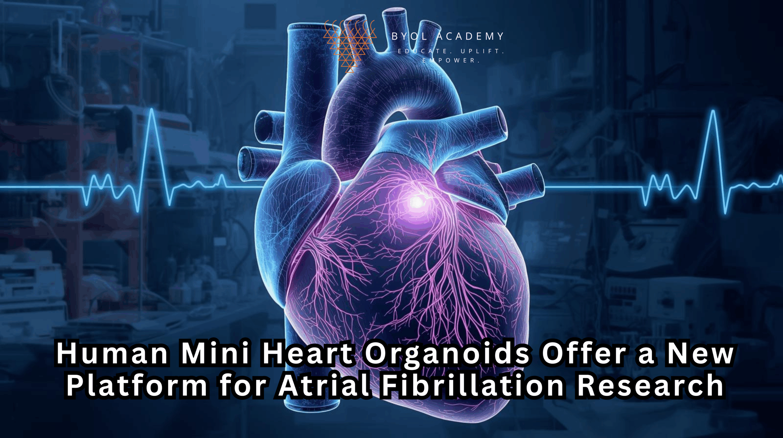
Recent advances in human stem cell biology and three-dimensional tissue engineering have begun to address this critical gap. The emergence of human heart organoids, self-organizing, multicellular cardiac tissues derived from pluripotent stem cells has opened new possibilities for modeling human heart development, disease, and therapeutic response in vitro. Building on this progress, scientists at Michigan State University have now developed vascularized, immune-competent human heart organoids that can be induced to exhibit atrial fibrillation–like electrical behavior. This innovation establishes, for the first time, a living human cardiac model that reproduces key features of AF pathophysiology, providing a transformative platform for mechanistic investigation and translational drug discovery.
Traditional AF research has relied heavily on animal models and simplified two-dimensional cardiomyocyte cultures. While valuable, these systems fail to capture critical features of the human heart, including three-dimensional architecture, multicellular interactions, human-specific electrophysiology, and the contribution of resident immune cells. As a result, many candidate therapies demonstrate promise preclinically but fail in human trials, perpetuating a decades-long stagnation in AF drug development.
Beginning in 2020, investigators led by Aitor Aguirre, PhD, associate professor of biomedical engineering and chief of the Division of Developmental and Stem Cell Biology at MSU’s Institute for Quantitative Health Science and Engineering, established protocols to generate self-organizing human heart organoids from donated pluripotent stem cells. These stem cells, capable of differentiating into multiple cardiac lineages, were guided through developmental programs that yield three-dimensional, beating cardiac tissues approximately the size of a lentil.
Crucially, the resulting organoids exhibit features far beyond simple cardiomyocyte aggregates. They develop chamber-like regions and complex vascular networks, including arteries, veins, and capillaries, enabling nutrient diffusion and sustained contractile activity. The organoids display robust, synchronized beating that is visible to the naked eye, reflecting functional electrical coupling and tissue-level organization comparable to native human myocardium .
A defining advance in the current study was the integration of innate immune cells, particularly macrophages, into the heart organoids. These cells are increasingly recognized as essential regulators of cardiac development, homeostasis, and pathological remodeling. Led by MSU physician-scientist trainee Colin O’Hern, the team demonstrated that macrophages within the organoids actively influence cardiac rhythm and maturation.
By experimentally inducing inflammatory signaling, a well-established contributor to AF in patients, the researchers triggered irregular, rapid beating patterns within the organoids that closely resemble atrial fibrillation. This represents the first human heart organoid model capable of recapitulating AF-like arrhythmia through defined biological mechanisms, rather than artificial electrical pacing or genetic manipulation alone. The findings were reported in Cell Stem Cell, underscoring their mechanistic and translational significance.
Beyond disease modeling, the organoids proved responsive to therapeutic intervention. When the investigators introduced an anti-inflammatory compound predicted to mitigate AF, the previously irregular beating patterns partially normalized, demonstrating restoration toward coordinated rhythm. This pharmacological reversibility confirms that the model is not merely descriptive but functionally predictive, enabling direct testing of candidate therapies on living human cardiac tissue.
This capability addresses a central limitation of prior AF research. Existing therapies largely target symptoms, such as rate control or rhythm suppression, rather than upstream disease drivers. The MSU organoid platform allows researchers to interrogate causal pathways, particularly the interplay between inflammation, immune cells, and cardiac electrophysiology, in a human-specific context.
In addition to modeling arrhythmia, the study yielded insights into human heart development. The researchers showed that long-lived, tissue-resident innate immune cells guide both structural maturation and rhythmic stability of the heart. These findings have implications beyond AF, informing our understanding of congenital heart disease, the most common class of birth defects worldwide.
The team further developed methods to “age” the organoids by exposing them to chronic inflammatory cues, generating tissue characteristics that more closely resemble adult myocardium. This advance overcomes a common limitation of stem-cell-derived cardiac models, which often resemble fetal or neonatal heart tissue and thus poorly predict adult disease behavior.
The broader implications of this work are substantial. The absence of reliable human AF models has been a major bottleneck in therapeutic development. By providing a human-based, multicellular, and physiologically relevant platform, these heart organoids have the potential to:
Consistent with the U.S. National Institutes of Health’s New Approach Methodologies (NAMs) initiative, this technology supports a shift toward more predictive, human-relevant preclinical testing frameworks.
Building on this foundational advance, MSU investigators are actively integrating these human heart organoids into pharmaceutical and biotechnology drug-development pipelines. Their immediate application lies in high-throughput compound screening, where candidate molecules can be evaluated simultaneously for anti-arrhythmic efficacy and cardiotoxic risk using living human cardiac tissue. This dual-assessment capability is expected to substantially improve the predictive accuracy of preclinical testing and reduce late-stage clinical trial failures.
Beyond population-level screening, a key long-term objective is the development of patient-specific heart organoids generated from an individual’s own cells. Such personalized models would allow investigators to examine inter-patient variability in disease mechanisms and drug responses, enabling precision-guided therapeutic selection. In parallel, ongoing refinements in vascularization, immune integration, and tissue maturation aim to produce organoids that more closely resemble adult human myocardium. Collectively, these advances lay the groundwork for future regenerative strategies, including the ambitious goal of generating transplant-ready cardiac tissues for repair or replacement of damaged myocardium.
The creation of immune-competent human heart organoids that reliably recapitulate atrial fibrillation constitutes a major conceptual and technological shift in cardiovascular research. By integrating stem cell biology, three-dimensional tissue engineering, immunology, and human-specific electrophysiology within a single experimental system, this platform overcomes critical limitations that have constrained AF research for decades. Importantly, it enables direct investigation of disease-driving mechanisms in living human heart tissue and provides a scalable, predictive framework for therapeutic discovery.
For atrial fibrillation, a condition with vast global prevalence yet minimal therapeutic innovation, these miniature, beating human hearts represent more than a technical achievement. They offer a realistic path toward mechanism-based treatments, safer drugs, and personalized care, potentially redefining how arrhythmias are studied and treated. In doing so, this work may mark the beginning of a new era in translational cardiac medicine, where human biology itself becomes the central engine of discovery.
Recent developments in regulatory science have marked a pivotal moment for hepatology and clinical research. The U.S. Food and Drug […]
Tobacco smoke has always been treated as something that fades once the air clears. Open a window, turn on a […]
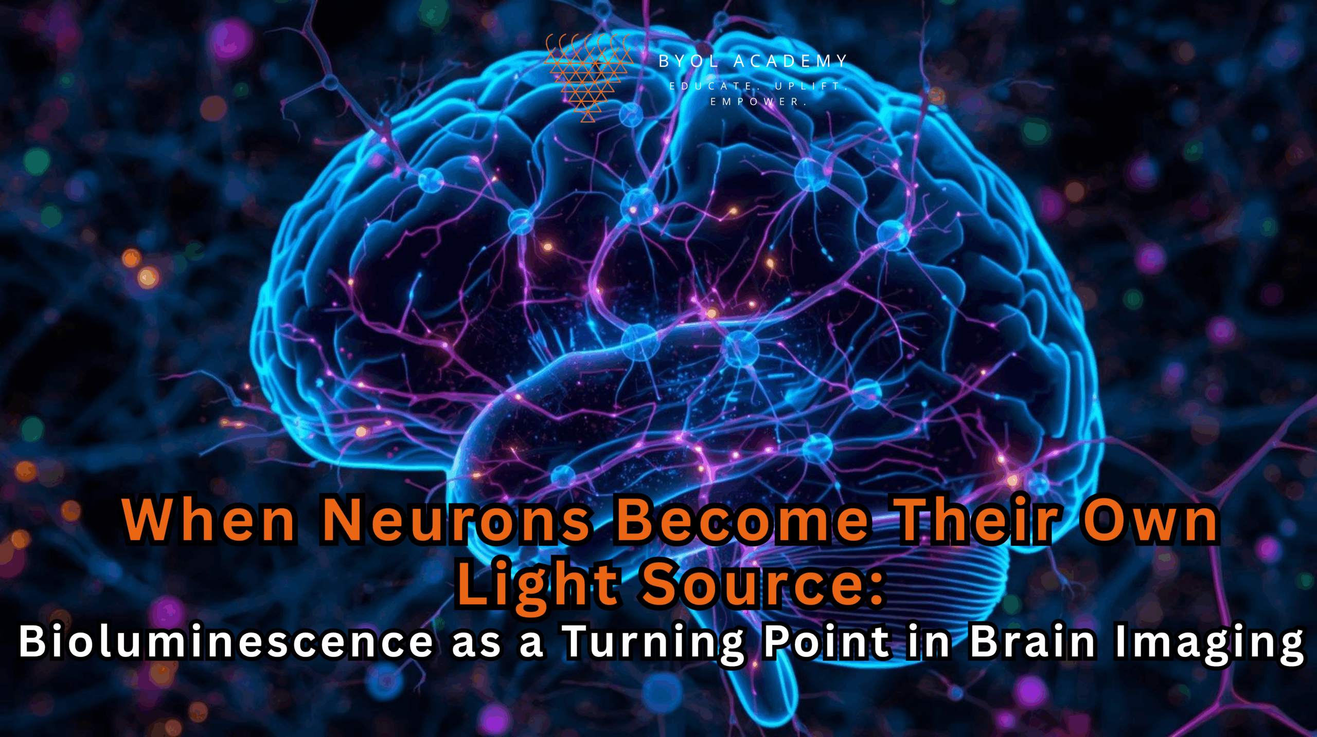
Neuroscience has long advanced by increasing the intensity of its gaze. To see deeper, faster, and with greater precision, researchers […]